|
MRI Modalities
|
|
BOLD fMRI :
 Blood oxygen level dependent (BOLD) responses to stimulus provocations represent the canonical method for non-invasive measurements of neural activation in real-time. While most stimulus presentation is achieved through the nose to take advantage of the primacy of the olfactory sensory modality in rodents, we also regularly use tactile stimulation and pharmacological agents (known as phMRI). Stimulation of other sensory modalities (e.g. sound) is under development. BOLD functional imaging is performed in awake animals using single-epoch stimulus provocation paradigms that allow us to image the unfolding pattern of activation across the entire brain in real time. Blood oxygen level dependent (BOLD) responses to stimulus provocations represent the canonical method for non-invasive measurements of neural activation in real-time. While most stimulus presentation is achieved through the nose to take advantage of the primacy of the olfactory sensory modality in rodents, we also regularly use tactile stimulation and pharmacological agents (known as phMRI). Stimulation of other sensory modalities (e.g. sound) is under development. BOLD functional imaging is performed in awake animals using single-epoch stimulus provocation paradigms that allow us to image the unfolding pattern of activation across the entire brain in real time.
Scans typically last 10-45 min.
Good for assessing stimulus-induced changes in neural activation across the entire brain, in real-time.
|
Quantitative anisotropy diffusion tensor imaging (QA-DTI)
 Quantitative anisotropy reflects the degree to which the diffusion of water (protons) varies in different directions. At a microscopic level there are many determinants of diffusion as the microarchitecture of brain parenchyma is composed of many components, including neurons and their axonal and dendritic fibers, glia, connective tissue, capillaries, and intracellular and extracellular water. Axonal properties and general microarchitecture within a voxel are the key determinants of diffusion anisotropy at a microscopic level, and the coherence of the axons in a voxel (i.e., are they parallel or crossing) is the key determinant of diffusion anisotropy at a macroscopic level. Quantitative anisotropy reflects the degree to which the diffusion of water (protons) varies in different directions. At a microscopic level there are many determinants of diffusion as the microarchitecture of brain parenchyma is composed of many components, including neurons and their axonal and dendritic fibers, glia, connective tissue, capillaries, and intracellular and extracellular water. Axonal properties and general microarchitecture within a voxel are the key determinants of diffusion anisotropy at a microscopic level, and the coherence of the axons in a voxel (i.e., are they parallel or crossing) is the key determinant of diffusion anisotropy at a macroscopic level.
Scans typically last 1 to 1.5 hrs and are performed on anesthetized animals.
Good for assessing changes in structure, regional microarchitecture.
|
Resting state functional connectivity (rfcMRI)
Resting state networks reflect neuronal baseline activity thought to measure low frequency fluctuations between functionally relevant brain areas. It is based on determining brain areas that demonstrate temporally correlated changes in BOLD signals, the most common used functional imaging modality. The default mode network refers to a resting state network with key brain areas, e.g. prefrontal and cingulate cortices, showing highly correlated brain activity and high metabolic demand. Functional connectivity is widely used in human imaging to monitor brain networks associated with normal perception, cognition, and emotion, and is also being used to identify abnormalities in connectivity associated with disordered function, such as addiction to drugs of abuse.
Scans typically last 10-20 min.
|
|
Volumetric anatomical analysis :
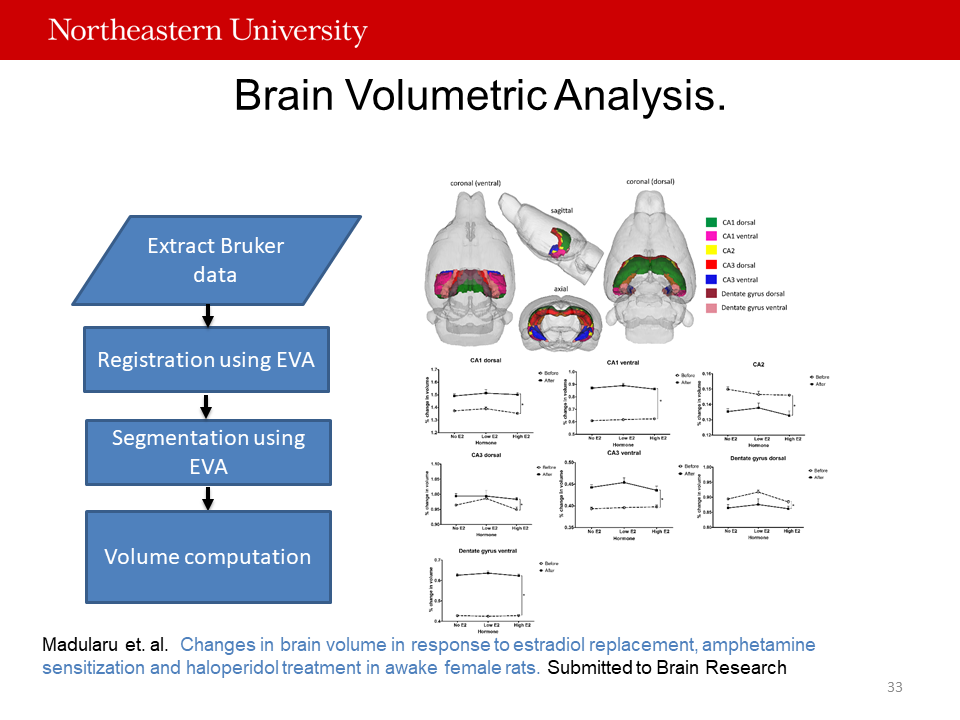 High-resolution anatomical scans can provide quantitative volumetric measurements of all regions included in each species’ 3D atlas. In this work we used predefined MRI Brain Atlas (Ekam Solutions LLC, Boston, MA) and registered the standard structural template image onto high resolution T2 weighted images for each individual subject using non-linear registration method implemented by Unix based software package Deformable Registration via Attribute Matching and Mutal-Saliency Weighting (DRAMMS). The atlas (image size 256x256x63) was then warped from the standard space into the subject image space (image size 256x256x40) using the deformation obtained from the above step using nearest-neighbor interpolation method. In the volumetric analysis, each brain region was therefore segmented and the volume values were extracted for all 171 ROIs, calculated by multiplying unit volume of voxel in mm3 by the number of voxels using in-house MATLAB script. To account for different brain sizes all the ROI volumes were normalized by dividing each ROI volume by total brain volume of that subject. High-resolution anatomical scans can provide quantitative volumetric measurements of all regions included in each species’ 3D atlas. In this work we used predefined MRI Brain Atlas (Ekam Solutions LLC, Boston, MA) and registered the standard structural template image onto high resolution T2 weighted images for each individual subject using non-linear registration method implemented by Unix based software package Deformable Registration via Attribute Matching and Mutal-Saliency Weighting (DRAMMS). The atlas (image size 256x256x63) was then warped from the standard space into the subject image space (image size 256x256x40) using the deformation obtained from the above step using nearest-neighbor interpolation method. In the volumetric analysis, each brain region was therefore segmented and the volume values were extracted for all 171 ROIs, calculated by multiplying unit volume of voxel in mm3 by the number of voxels using in-house MATLAB script. To account for different brain sizes all the ROI volumes were normalized by dividing each ROI volume by total brain volume of that subject.
|
|
T1 & T2 Maps :
 A workflow based on the ratio between standardized T1-weighted and T2-weighted magnetic resonance images serves as a new tool to image healthy brains. This technique, examining the utility “mapping” of the longitudinal and/or transverse (T1 and T2) relaxation times in the context of neurologic disease have demonstrated variations in T1 and T2 within specific brain regions within autism, schizophrenia, epilepsy, Parkinson’s, multiple sclerosis, and a host of other disorders. A workflow based on the ratio between standardized T1-weighted and T2-weighted magnetic resonance images serves as a new tool to image healthy brains. This technique, examining the utility “mapping” of the longitudinal and/or transverse (T1 and T2) relaxation times in the context of neurologic disease have demonstrated variations in T1 and T2 within specific brain regions within autism, schizophrenia, epilepsy, Parkinson’s, multiple sclerosis, and a host of other disorders.
|
|
Manganese-enhanced MRI (MEMRI) :
Manganese serves as an essential for proper physiological functioning, as well as a calcium analog and can be involved in neuron firing (voltage gated channels). These properties of Mn2+ make it a useful contrast agent for anatomical and functional imaging in multiple systems, and can be used for enhancement of the brain cytoarchitecture for anatomical studies. MEMRI has been used to trace neuronal pathways, define morphological boundaries, and study connectivity in morphological and functional imaging studies.
Furthermore, given that Mn2+ is transported by microtubule-dependent axonal transport and can cross synapses to reach postsynaptic neurons, MEMRI has been used as a neuronal tract tracer for several neuronal pathways including the visual, olfactory, and somatosensory pathways, in a variety of animal models, such as mice, rats, monkeys, and birds. Currently, MEMRI has three primary applications in biological systems: contrast enhancement for anatomical detail, activity-dependent assessment and tracing of neuronal connections or tract tracing.
|
|
Magnetic Resonance Spectroscopy :
Magnetic Resonance Spectroscopy (MRS) captures information of concentration of brain metabolites such as NAA, Cho, Cr and lactate in the tissue examined. This study is good for evaluating CNS disorders, identifying specific patterns of metabolites, and identifying high grade from low grade brain tumors (MRS can compare the chemical composition of normal brain tissue with abnormal tumor tissue).
|