Salamanders are capable of regenerating spinal cord tissue following transections, crushes, or amputations. Click here to see a time-lapse movie of axolotl tail regeneration over 45 days.
tail regeneration
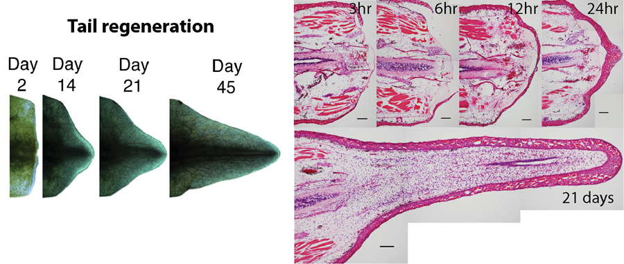
The figure below shows the external morphology of tail regeneration as well as the internal changes that are characteristic to appendage regeneration. The tissue sections on the right are coronal (viewed from the back of the animal downward) to show the sequence of events that will lead to complete regeneration. Notice the spinal cord in the middle of each section. Also notice that the wound epidermis has migrated over the exposed stump by 12 hours post amputation to close off the injury from the external environment. Cells near the injury site then start to proliferate to generate a structure called a blastema. Over the next several weeks, the blastema grows in a similar manner as limb regeneration except a new spinal cord is present in the middle of the tail blastema (hollow portion in 21 day histological section).
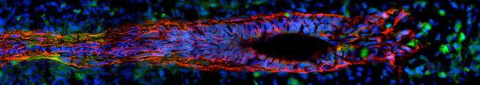
Over time, a spinal cord is completely regenerated, including new nerve cells and the supporting cells that are necessary for proper motor control and sensory input. The image below shows the cells responsible for this process within a tail blastema. The red cells are neural progenitor cells that will eventually differentiate into new neurons, while the green labels inflammatory cells and the blue labels nuclei. Our research identifies, characterizes, and tests the function of genes that are expressed during spinal cord regeneration in order to understand the process at the cellular and molecular level. The hypothesis is that the cell types and gene expression patterns specific to the salamander compared to non-regenerating animals endow them with regenerative abilities.
STUDIES ON SPINAL CORD REGENERATION
Investigating Nrg1 Signaling in the Regenerating Axolotl Spinal Cord Using Multiplexed FISH
Freitas, P. D., Lovely, A. M., & Monaghan, J. R. (2019). Developmental neurobiology, 79(5), 453-467.
Spinal Cord Regeneration in Amphibians: A Historical Perspective
Freitas, P. D., Yandulskaya, A. S., & Monaghan, J. R. (2019). Developmental neurobiology, 79(5), 437-452.
Expression pattern of Nogo-A, MAG, and NgR in regenerating urodele spinal cord
Hiu SP, Monaghan JR, Voss SR, Ghosh S.
Dev Dynamincs. 2013 242(7):847-860.
Early gene expression during natural spinal cord regeneration in the salamander Ambystoma mexicanum.
Monaghan JR, Walker JA, Beachy CK, and Voss SR.
J Neurochem. 2007 101(1):27-40.